 CONFERENCE
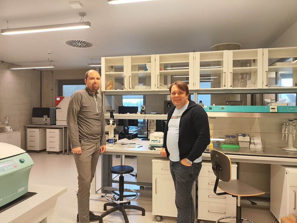
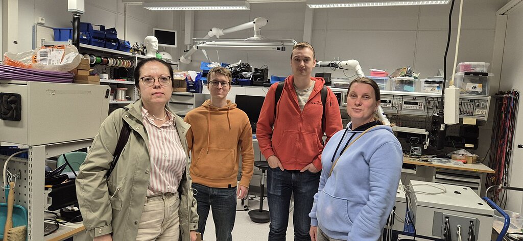
From 25 November to 8 January 2024 Dr. Sergiy Kyrylenko visited Department of Technical Physics, University of Eastern Finland in Kuopio (Itä-Suomen yliopisto), Finland.
The visit started with intensive exchange of knowledge in the truly interdisciplinary fields. The objective of the visit was to evaluate if the technique of electric impedance tomography could be applied to study conductivity of the polymer-MXene membranes. As a result, the experimental setup was brainstormed and the design of the experiment was put together. To the best of our knowledge, that was the first approach to evaluate the spatial distribution of the conductivity of the polymer-MXene membranes.
Despite the extreme winter weather conditions, the visit was a complete success. During the visit the secondee received training in non-destructive electrical imaging techniques, while the host Department obtained new directions in working with MXenes and other nanoparticles. The research results of the visit will be utilised in the further investigations of the polymer-MXene membranes in the in-vivo experimental models.

From 21 February to 20 April 2024, Dr. Sergiy Kyrylenko from the Sumy State University conducted a research secondment to Universidade Estadual de Campinas (Unicamp). During the secondment, the electro-conductive polycaprolactone (PCL) electrospun nanofibrous membranes with Ti3C2 MXene surface layers, produced by the Sumy State University in collaboration with Materials Research Centre – MRC, were first tested in the conditions in-vivo to support neuronal regeneration in animal model of sciatic nerve injury in rats. To obtain more reliable and reproducible results, the similar membranes were produced also at the University of Latvia in Riga, Latvia.
At the Unicamp, together with Prof. Alexandre Oliveira and his team, a series of experiments were conducted to investigate how neuronal conduits, made out of MXene covered PCL nanofibrous membranes, could be used in design of neuronal conduits to support regeneration of sciatic nerves in the animal model of neuronal injury in rats. For this, the sciatic nerve was dissected and sutured by neurorrhaphy in conjunction with axon fusion technique. Functional recovery was monitored by CatWalk system during 4 weeks post operation. After that, the neuronal fibers together with the conduits and surrounding tissues were examined and prepared for transmission electron microscopy (TEM) investigations.
As a result, while the data processing is still in progress, it was showed that the functional regeneration occurred much faster than after the traditional neurorrhaphy. Importantly, it was also observed that the new conduits did not induce adverse accumulation of connective tissues at the site of treatment. Currently, experiments are in progress to show the microstructure of the axonal fusion and the state of the MXenes and the PCL nanofibers after 4 weeks in the animal’s body. In addition, currently a second series of experiments are planned to investigate functional recovery during 8 weeks post operation, as well as histological examination of the treatment sites.
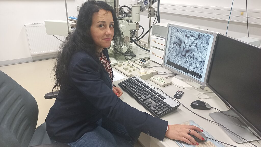
From 11 to 25 April 2024 Dr. Volodymyr Deineka visited the Vilniaus universitetas, Vilnius, Lithuania.
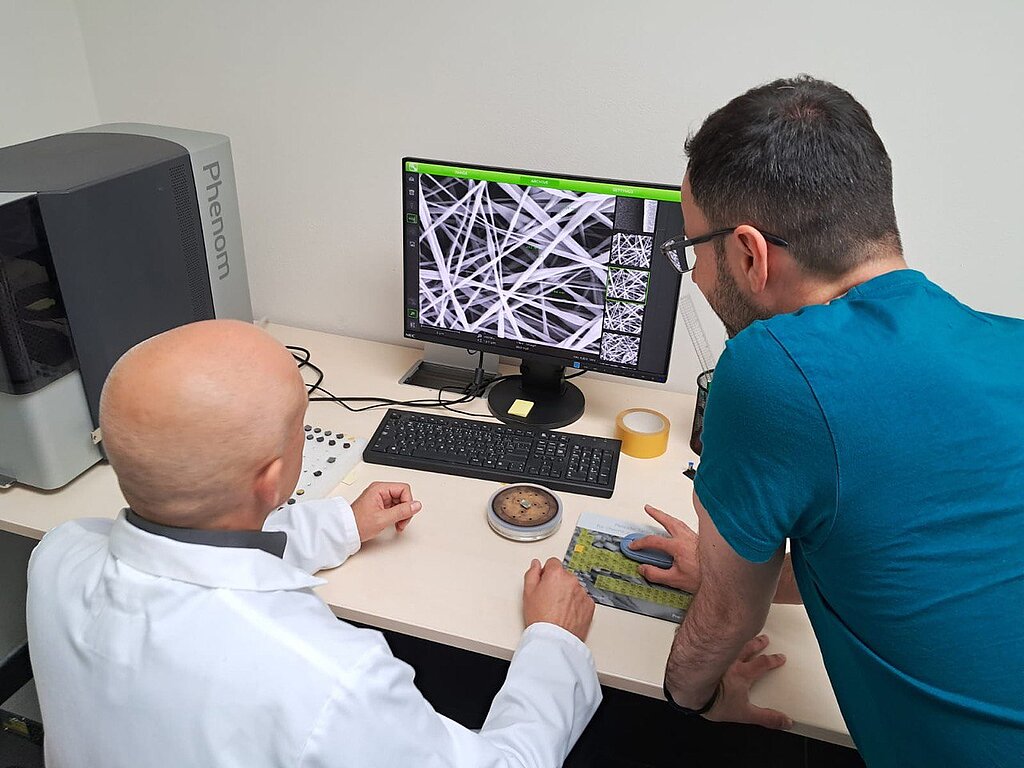
The visit aimed to conduct a structural description of the Ti3C2 and Nb2C Mxenes. Data between these two types of MXenes will be compared, and patterns of characteristics associated with the size of nanoparticles will be determined. During the implementation, Dr. Deineka received intensive training on working with SEM. The Vilniaus universitetas staff demonstrated best practices in studying nanomaterials.
After obtaining the structural characterisation results, the data were discussed to select the best samples to be coupled to the nanofibrous 3D membranes. Dr. Deineka demonstrated different types of polymer membranes obtained by electrospinning and spoke about the features of producing these materials. As a result of the discussion, the optimal parameters of the membrane were selected as a scaffold for applying Mexene.
The results of the visiting studies will be used in further studies of polymer-MXene membranes on experimental models in vitro.

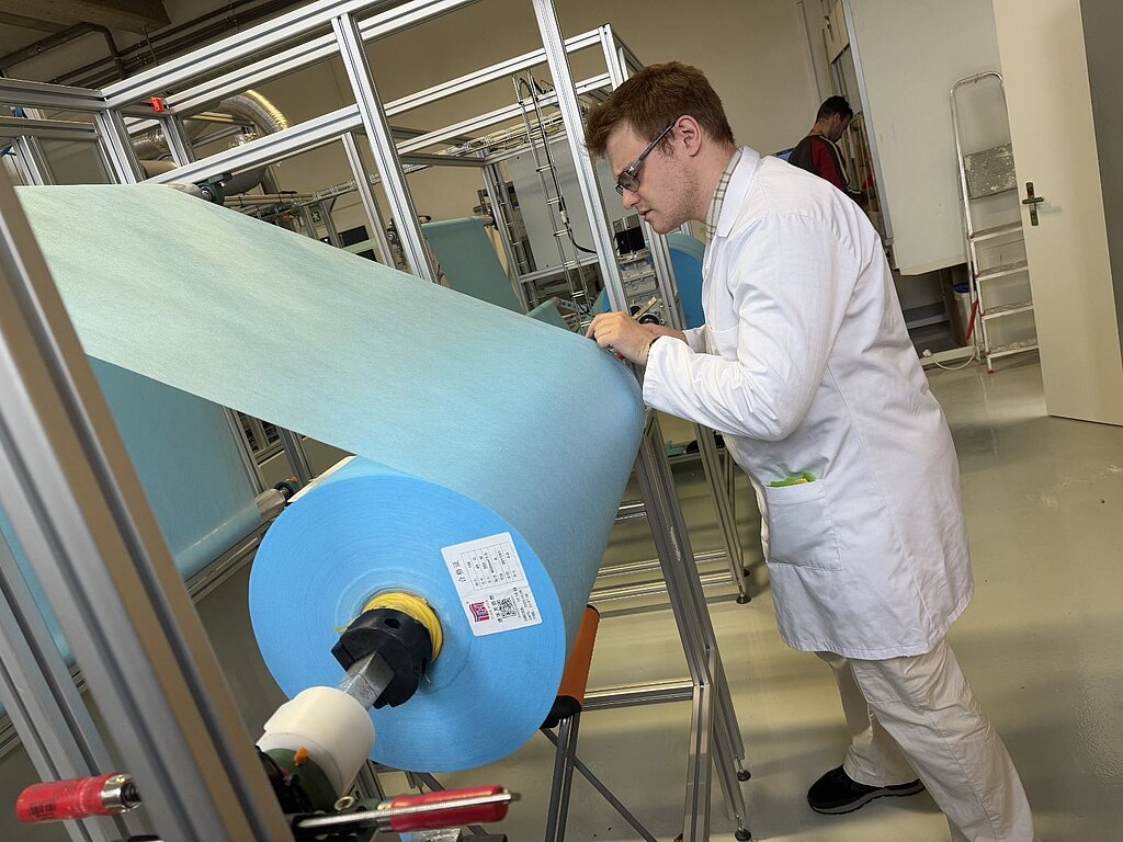
From 8 April to 7 August 2024 Vipin Kumar Veeramuthu visited University of Latvia.
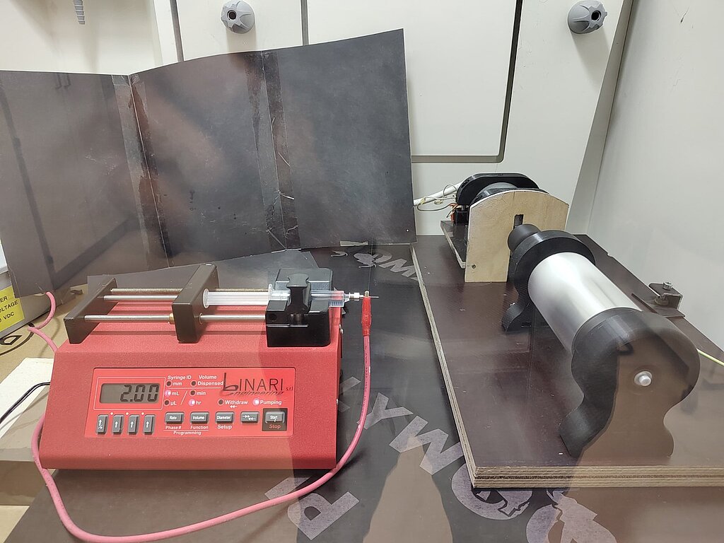
During a four-month secondment at the University of Latvia, the assembly and commissioning of an electrospinning unit was successfully completed. This included the integration of key components such as pumps and collectors, all of which were sourced directly from LINARI ENGINEERING SRL, Italy. Rigorous testing ensured that the system operated seamlessly and achieved optimum performance.
This was followed by the selection of specific solution parameters, including solvent types, concentrations and nanofiber electrospinning processing conditions. These parameters were carefully selected to develop polymer solutions using polycaprolactone (PCL), polylactic acid (PLA) and chitosan tailored for the production of 3D electrospun membranes. Using conventional, multi-needle and coaxial electrospinning techniques, the desired membrane morphology and properties were achieved through careful manipulation of solution parameters and electrospinning conditions.
Finally, after numerous experiments and analyses, the 3D electrospun PCL membranes with the desired properties were prepared and can be used for experiments with MXenes. Other polymers were still being optimized to obtain perfect membranes and this will be continued in the next period of secondment.
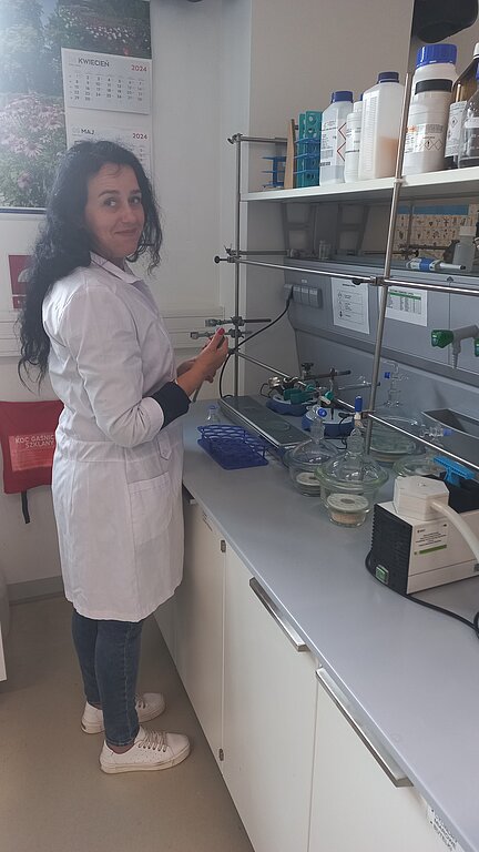
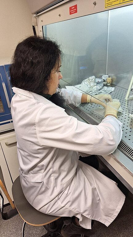
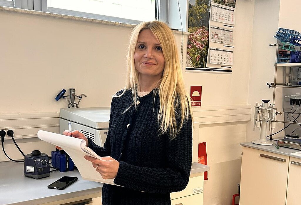
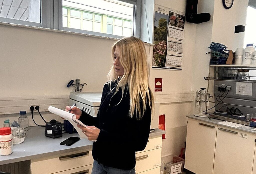
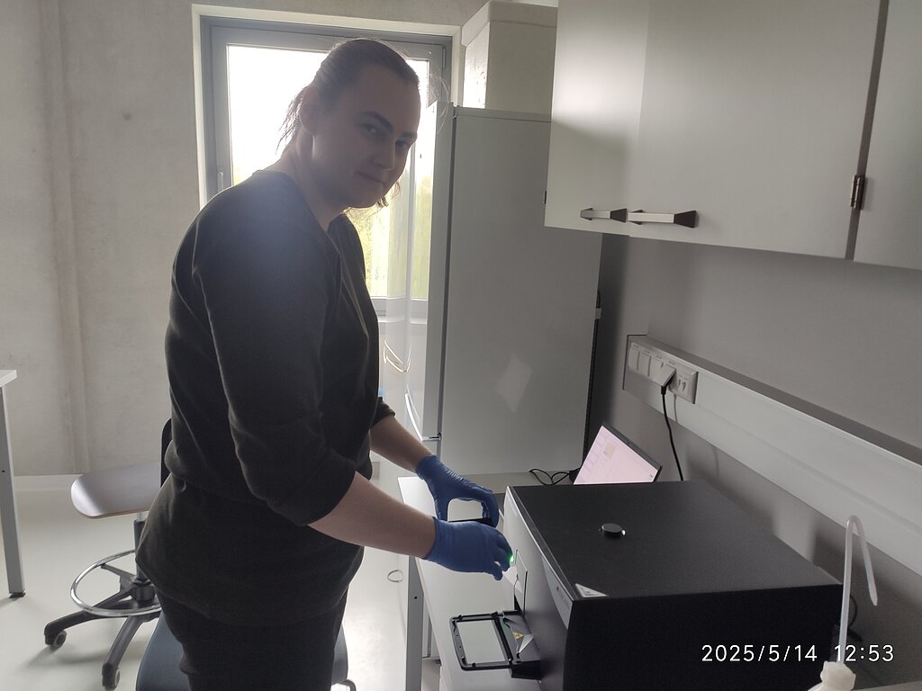
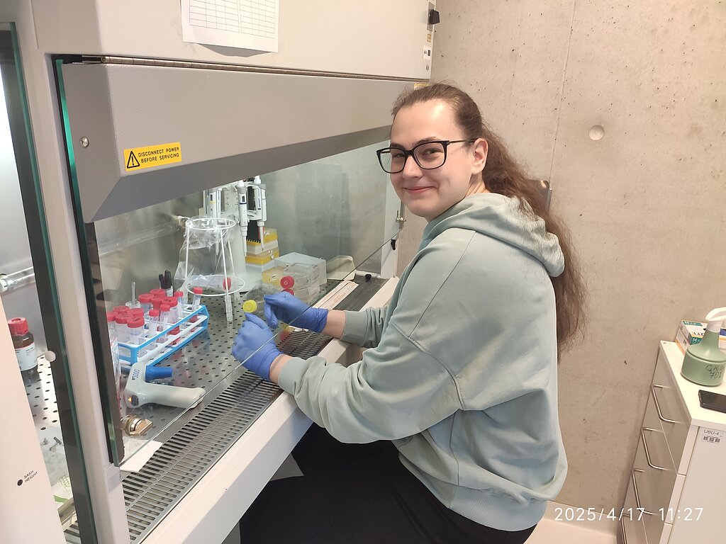
From 22 April to 21 May 2024, Ph.D. student Yevheniia Husak visited the THE ADAM MICKIEWICZ UNIVERSITY, Poznan, Poland.
During my visit, I conducted a series of comprehensive training sessions focused on laboratory safety protocols. The primary objective of this visit was to develop electro-conductive polymeric 3D scaffolds. My involvement included several key activities. Firstly, I meticulously prepared the PCL electrospun membranes with incorporated MXene at different concentrations to assess its impact on the scaffolds' conductive properties. Subsequently, I engaged in detailed morphological testing of the resultant membranes using a scanning electron microscope (SEM). This analysis was crucial to evaluate the surface structure, uniformity of MXene attachment, and the depth of MXene penetration within the membranes. Following the SEM analysis, I conducted further characterization of the membranes using Fourier-transform infrared spectroscopy (FTIR) and Raman spectroscopy. These techniques were essential to understand the chemical interactions and structural integrity of the MXene-loaded membranes. FTIR provided insights into the functional groups present and any chemical modifications, while Raman spectroscopy offered detailed information about the molecular vibrations and crystallinity of the materials.
Through these meticulous processes, we aimed to develop high-performance, electro-conductive polymeric 3D scaffolds with potential applications in tissue engineering and other biomedical fields. The comprehensive testing and characterization ensured that the scaffolds met the desired specifications and performance criteria.
The visit was carried out on research project Horizon Europe MSCA-SE project “Electro-conductive polymeric 3D scaffolds as novel strategies for biomedical applications”.


Prof. Aku Seppänen from the University of Eastern Finland (UEF, Finland) visited the Institute of Electronics and Computer Science (EDI, Latvia) from May 1st, 2024 to May 15th, 2024.
The visit in the frame of the Horizon Europe MSCA-SE project “Electro-conductive Polymeric 3D Scaffolds as Novel Strategies for Biomedical Applications” (ESCULAPE) was devoted to MXenes testing including monitoring of MXenes conductivities in varying humidity conditions, effect of materials used in MXenes to its conductivity (e.g. use of Niobium (Nb) or Vanadium (V) instead of Titanium), the effect of Ti flake size in the solution to the MXenes conductivity and monitoring of the MXenes degradation process.
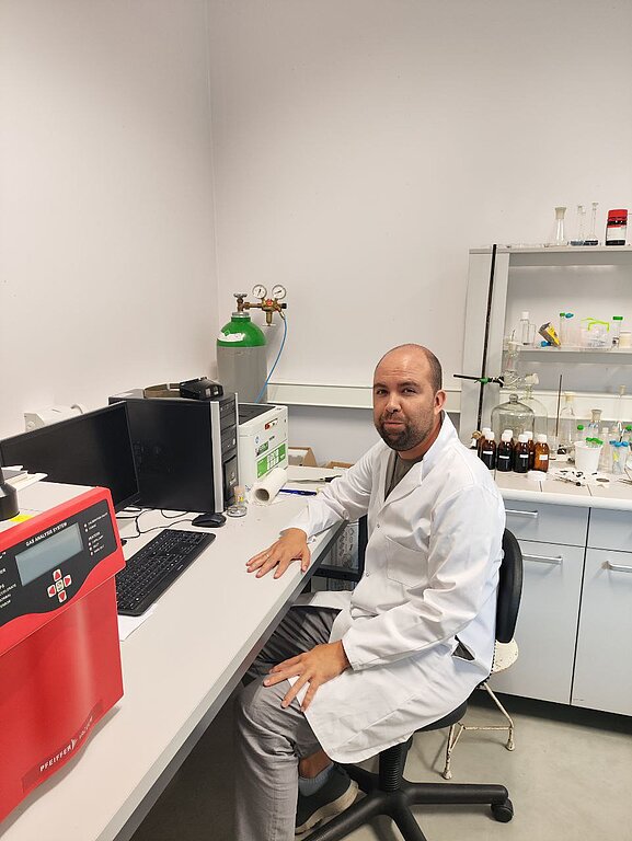
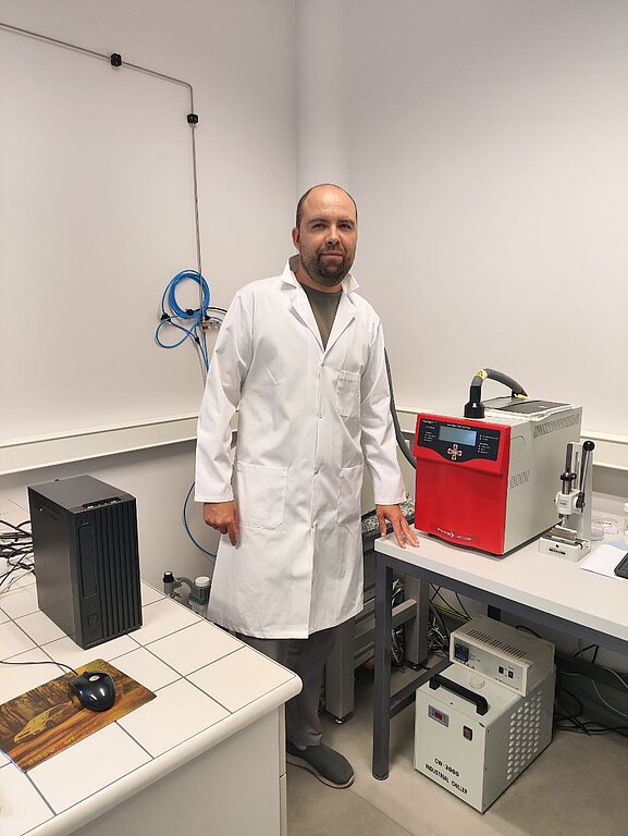
Anton Popov visited the NanoBioMedical Center, AMU (Poznan, Poland) from June 9, 2024 to July 8, 2024.
During the secondment, work on the synthesis and characterization of MXenes Ti3C2Tx was carried out. In addition, the deposition of MXenes by different methods on the electrode surface was studied. Electrochemical measurements were used to investigate the electrodes modified with the Ti3C2Tx MXenes layer. The optimal deposition parameters of MXenes layers were selected.
Dr. Anton Popov imparted knowledge on the synthesis and characterization of MXenes. Intensive training on spray coating techniques for the deposition of MXenes layer on different surfaces was carried out. The results obtained will be useful for the future deposition of MXenes on polymer membranes.
From June 9th to August 8th, 2024, PhD student Katazyna Blazevic visited Uniwersytet im. Adama Mickiewicza w Poznaniu.
The purpose of the secondment was the development and optimization of MXenes nanocomposites using various coating techniques, followed by optical and electrochemical characterization.
During the secondment, Katazyna Blazevic learned about layer formation by spray coating technique for MXenes and the MXene-TiO2 composite. To assess the deposition of nanocomposite layers on the surface, the photoelectric characteristics of these layers were examined. The characterization of the produced layers was performed by employing photoelectrochemical, optical, SEM, and EDX techniques.
Optimal conditions for layer formation by the spray coating technique were determined and achieved. Promising results were obtained, showing a substantial increase in photocatalytic activity compared to pure TiO2 nanowires.
In future works, MXene nanocomposite deposition by spray coating technique can be used to deposit such nanocomposites on the polymeric membranes.
Aleksi Kuivisto from the University of Eastern Finland (Itä-Suomen yliopisto) visited the Institute of Electronics and Computer Science (EDI) from July 1st, 2024, to July 30th, 2024.
During a one-month visit in the frame of the Horizon Europe MSCA-SE project, “Electro-conductive Polymeric 3D Scaffolds as Novel Strategies for Biomedical Applications” (ESCULAPE), measurement of the electrical conductivities of MXene-based electrospun scaffolds was carried out. As the first step, experiments to study the electrical conductivity development of MXenes after dip coatings. Here, the technique developed in UEF, Electrical impedance tomography (EIT), was used in EDI to overcome preliminary tests conductivity evaluation challenges based on MXenes inhomogeneous after scaffold coating.
Dr. Mykola Pavlenko and PhD student Andrii Lys from AMU (Poznan, Poland) visited VU (Vilnius, Lithuania) from August 5th to September 4th, 2024.
During the visit in the frame of the Horizon Europe MSCA-SE project “Electro-conductive Polymeric 3D Scaffolds as Novel Strategies for Biomedical Applications” (ESCULAPE), Dr. Mykola Pavlenko and PhD student Andrii Lys received scientific training in the synthesis of noble metal nanoparticles and conducting polymer layers. Deposition of MXene-based electrodes for subsequent electrochemical analysis involved the preparation and optimization of MXene-based electrodes to ensure their effectiveness and stability across various applications.
During a six-month secondment (Vipin Kumar Veeramuthu) at the University of Latvia, the electrospinning setup was modified to optimize the fiber arrangement within the 3D membrane. The primary modifications involved adjusting the collector dimensions and rotation speed to achieve the desired structure.
Electrospun polycaprolactone (PCL) membranes with well-defined structures, as confirmed by scanning electron microscopy (SEM), were further developed by incorporating MXene particles into the spinning solution. The presence of MXenes on the nanofibrous membranes was subsequently analyzed. Membranes with aligned fiber structures were successfully fabricated using various electrospinning techniques and characterized through SEM imaging.
Additionally, polymers such as PCL and polyethylene oxide (PEO) were individually combined with chitosan to produce electrospun membranes. However, achieving the optimal membrane structure remains challenging and requires further optimization. Several membranes with promising structural integrity have been successfully prepared and will be used in subsequent experiments.
During her secondment at INSERM from 2nd September 2024 to 21st December 2024, Maria João Ferreira from Biofabics gained invaluable experience in human induced pluripotent stem cell (hiPSC) culture and differentiation. The visit allowed her to delve into 2D and 3D differentiation protocols, focusing on the generation of hiPSC-derived cardiomyocytes.
Maria João was introduced to the 2D differentiation process, where she worked on establishing protocols for differentiating hiPSCs into functional cardiomyocytes. Additionally, she developed 3D cardiac organoids by applying a specialized differentiation protocol designed for 3D culture conditions, which is an essential model for more accurate cardiac tissue engineering.
One of the most impactful aspects of her visit was the exposure to electrophysiology techniques, particularly Microelectrode Array (MEA) recordings. These techniques allowed her to study the electrical activity of hiPSC-derived cardiomyocytes and organoids, providing a deeper understanding of cardiac cell function.
The interdisciplinary nature of the team at INSERM allowed Maria João to work alongside experts from various fields, broadening her scientific knowledge and expanding her skills. She had the opportunity to explore a wide range of methodologies and research topics, which enriched her overall experience.

The secondment began with Ines Marques attendance at the IEEE NAP 2024 Conference, held in Riga from September 8 to 13. This conference brought together leading experts in nanomaterials and their applications, offering valuable insights into cutting-edge research and providing opportunities to establish professional connections within the field.
At the University of Latvia, the main experiment conducted aimed to evaluate how MXenes influenced the expression of specific genes in different cell types exposed to varying concentrations of these materials. In addition, she took the opportunity to deepen her understanding of the electrospinning technique, which will be essential for future projects.
Beyond laboratory work, she attended seminars and participated in team-building activities, which provided important insights and opportunities to connect meaningfully with colleagues.
This secondment was a rewarding experience that enabled her to develop her research skills, contribute to innovative projects, and achieve personal and professional growth in an inspiring environment.
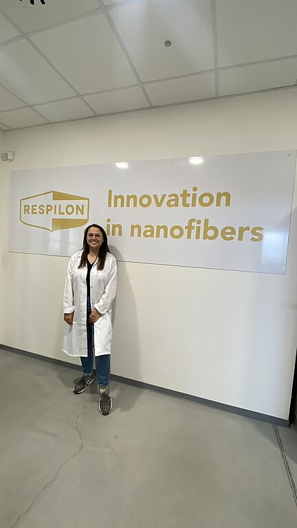
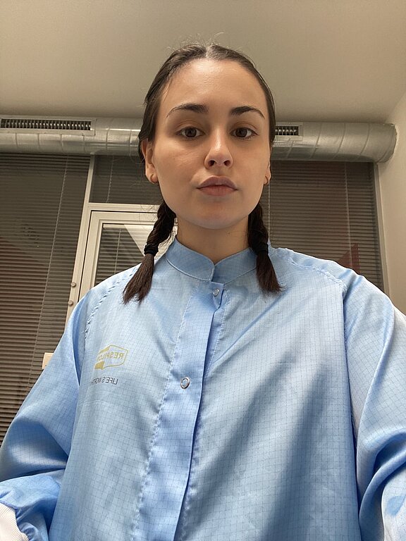
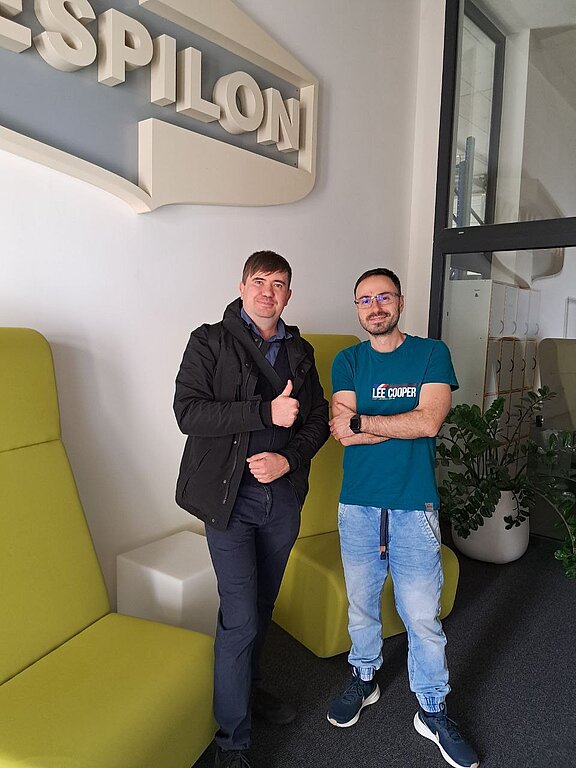
Viktorija Liustrovaite from Vilnius University (Lithuania) visited RESPILON in Czechia from September 8, 2024, to October 7, 2024.
During her secondment, Viktorija received hands-on training in preparing MXene solutions and depositing them onto nanofibers. She was trained in electrospinning techniques, focusing on the fabrication of PCL nanofibers and the optimization of processing parameters. Additionally, she gained experience in using SEM to characterize the morphology and surface structure of the coated nanofibers.

From 16th September to 15th November 2024, the secondee, Marina Sapauskiene, visited Respilon for a two-month secondment.
The main goal of the secondment was the preparation and characterization of MXene-coated nanofibers using electrospinning and direct-coating techniques. During this time, various concentrations of ethanolic and aqueous MXene solutions were prepared to optimize coating processes. The characterization of MXene-coated nanofibers was performed using scanning electron microscopy (SEM) and voltage testing. These methods allowed for the evaluation of morphological properties and the effectiveness of MXene deposition on nanofiber surfaces.
Regular training sessions were held during the secondment, providing expertise in the preparation of MXene solutions and nanofiber coating methods. The secondee also received hands-on training in electrospinning, covering all steps from polymer solution preparation to fiber production. In addition, detailed instruction on SEM techniques, including sample preparation and image acquisition, was provided.
Future research will focus on improving nanofiber coating techniques by developing multilayered coatings to enhance material properties. The long-term stability of the coated nanofibers will be evaluated, including durability tests under repeated washing cycles. Efforts will also be directed towards producing biocompatible nanofibers coated with MXenes for applications in fields requiring biocompatibility."
During the recent activities (Oleksandr Solodovnyk), significant progress was made in evaluating MXenes synthesized and delaminated using various methods. Collaborative efforts focused on analyzing the structural and compositional attributes of MXenes to optimize their performance for advanced applications.
The assessment involved implementing a comprehensive characterization protocol to ensure a thorough understanding of the material properties. Structural characterization techniques, including scanning electron microscopy (SEM), X-ray diffraction (XRD), X-ray photoelectron spectroscopy (XPS), energy-dispersive X-ray analysis (EDX), and ultraviolet-visible spectroscopy (UV), were utilized to analyze the synthesized MXenes. These characterizations are integral to determining the impact of synthesis and delamination modes on the structural integrity and functional capabilities of MXenes.
Future work will involve correlating the characterization results with specific synthesis and delamination parameters to refine the production processes. Additional efforts will also aim at extending the characterization protocols to include thermal and mechanical property evaluations. This systematic approach will pave the way for developing highly tailored MXenes suited for targeted applications.
During this secondment Beatriz Carneiro, the study explored MXene coating on various nanofibers, assessing stability, retention of properties, and washing effects, with a focus on conductivity. Activities included sample centrifugation, solvent purification, vacuum-assisted MXene coating on Respilon nanofibers, and ethanol washing. SEM and conductivity measurements were used for evaluation.
Results showed uniform MXene coating by visual inspection, though precision could improve with specialized equipment. Centrifugation was ineffective for washing MXenes in dispersion, and post-coating washing caused nanofiber tearing and MXene loss. Further studies should explore alternative washing methods and their impact on MXene and nanofiber properties.During this secondment, the study explored MXene coating on various nanofibers, assessing stability, retention of properties, and washing effects, with a focus on conductivity. Activities included sample centrifugation, solvent purification, vacuum-assisted MXene coating on Respilon nanofibers, and ethanol washing. SEM and conductivity measurements were used for evaluation.
Results showed uniform MXene coating by visual inspection, though precision could improve with specialized equipment. Centrifugation was ineffective for washing MXenes in dispersion, and post-coating washing caused nanofiber tearing and MXene loss. Further studies should explore alternative washing methods and their impact on MXene and nanofiber properties.

During a two-month secondment Viktoriia Kazban at the University of Latvia, solution parameters such as solvent types, concentrations, and electrospinning conditions were optimized to develop polymer solutions from polycaprolactone (PCL), polylactic acid (PLA), and chitosan for producing 3D electrospun membranes. Using conventional, multi-needle, and coaxial electrospinning, the desired membrane properties were achieved through precise parameter adjustments.
After extensive experiments, 3D electrospun PCL membranes with the required properties were prepared for MXene-related tests. Optimization of other polymers is ongoing and will continue in the next project phase.
This work aims to develop solutions for tissue engineering, cardiac and nerve regeneration, iPSC research, and wearable electronics.
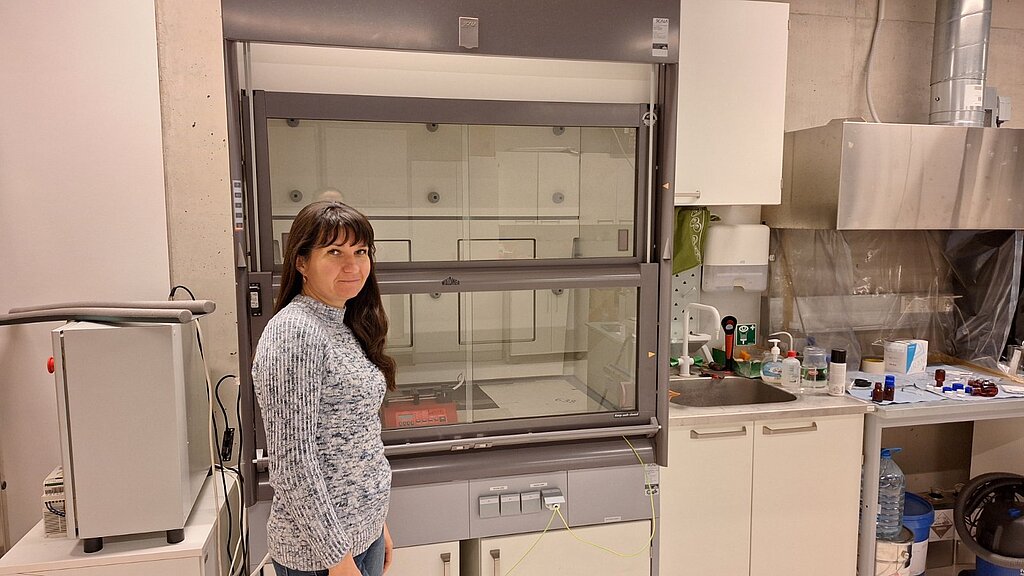
From November 1th to November 30th, 2024, Viktoriia Holubnycha from SSU (Sumy, Ukraine) visited LU (Riga, Latvia).
In the frame of the secondment period, substantial progress was achieved in the development of porous 3D electrospun membranes using a variety of polymers and polymer blends, including PCL, PLA, and chitosan. Efforts were focused on optimizing the electrospinning process to produce membranes with precisely controlled porosity and enhanced structural integrity, making them suitable for advanced biomedical and technological applications. These studies aim to bridge the fields of materials science, regenerative medicine, and wearable technology, contributing to the development of next-generation biomedical devices and therapies.

During the secondment (Alexander Tamashevski), Ti₃C₂-based MXene nanosheets were synthesized using a top-down chemical etching method from bulk Ti₃AlC₂ ceramics with hydrofluoric acid. Ultrasonic delamination was performed using tannic acid-based polyphenols, while TPAOH was intercalated to introduce hydroxyl groups (MXene-OH). This modification facilitated the covalent attachment of biodegradable polymers such as polycaprolactone (PCL) and PEtOx. Additionally, MXene nanosheets were further functionalized with PCL via anionic polymerization using tin (II) octoate as a co-initiator and MXene-OH as the reaction initiator.
Furthermore, the secondee underwent specialized training in MXene synthesis and surface modification, significantly enhancing their expertise in developing 3D matrices with improved structural and mechanical properties.
During the secondment visit(Mykola Pavlenko,Andriy Lys) , a comprehensive scientific training program was conducted, focusing on factory-scale nanofiber production. Company representatives provided demonstrations of their production processes and discussed common challenges encountered in electrospinning. This training offered unique insights into the behavior of various polymers within polymeric solutions. Multiple dissolvers were tested to highlight differences in polymer solubility and behavior during electrospinning.
In addition, several test polymeric solutions containing MXenes were examined through electrospinning to determine the optimal parameters for the future design and development of MXene-based 3D membranes. Host researchers provided invaluable supervision, advice, and hands-on opportunities to mix different polymer solutions and assess their electrospinning potential.
During the visit Yevheniia Husak, significant progress was achieved in advancing the alcohol-assisted deposition method for integrating MXene into PCL electrospun membranes.
These results are especially critical for regenerative medicine, where reliable and biocompatible scaffolds are essential for success. The MXene-modified PCL membranes developed during this visit are well-aligned with the needs of conductive tissue engineering, particularly in challenging applications such as cardiac and neural regeneration.
Building on these achievements, future efforts will focus on evaluating the performance of the MXene-modified PCL membranes in biological systems through in-vitro and in-vivo testing. This will provide critical insights into their potential for clinical use. Furthermore, optimization of the alcohol-based solution composition will be explored to enhance deposition efficiency and ensure the long-term stability of the modified membranes. These steps aim to refine the technology and expand its applications in tissue engineering and regenerative medicine.
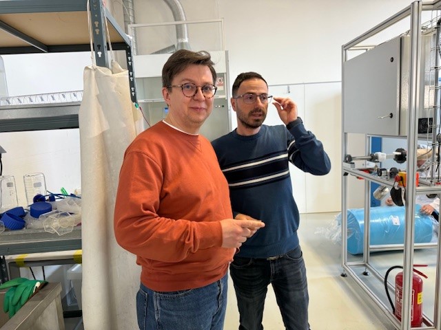
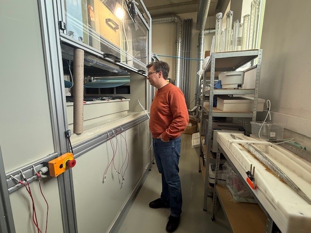
From November 12 to November 26, 2024, Dr. Maksym Pogorielov and From November 11 to November 25, 2024 Dr. Volodymyr Deineka visited RESPILON MEMBRANES S.R.O., Prague, Chech Republic.
This visit aimed to study the features of industrial electrospinning and work with two independent high-voltage sources.The production of fibers with incorporated Mexenes nanolaminates was studied. Several working solutions of biopolymers with different concentrations of Mexenes were studied, and work was carried out to adapt the electrospinning modes. During the implementation, Dr. Deineka underwent intensive training in preparing and characterizing polymer solutions for electrospinning, as well as the effect of viscosity and electroconductivity of the solution on its ability to electrospinning. Dr. Pogorielov studied the dependence of changes in membrane electrical conductivity on the concentration of MXenes. The employees of RESPILON MEMBRANES S.R.O. demonstrated the best practices in manufacturing a clean line to produce electrospun membranes. Dr. Pogorielov shared the features of working with MXenes and their storage. The obtained PCL-MXene membranes will be studied for cytotoxicity and biodegradation. The obtained data will be used to prepare a joint publication upon completing all studies.
During the secondment period from December 18 to 22, Oleksiy Gogotsi and Veronika Zahorodna from MRC visited LU, where a master class on the preparation, handling, and characterization of MXene material samples was conducted. A set of MXene samples at various concentrations was prepared for upcoming biological experiments. Additionally, a research meeting was held to discuss the latest progress and experimental findings.
As part of the dissemination activities, a workshop focused on the biological applications of MXene materials was organized at LU, involving researchers from LU and partner institutions. Project results and updates were presented and discussed among the participants, enhancing visibility and fostering collaboration across institutions. Training sessions on MXene handling and sample preparation were also provided, covering key protocols for safe and effective material processing, including stabilization techniques and concentration management.
In the long-term perspective, the outcomes of this secondment will contribute to ongoing research in the fields of biocompatibility and biomedical applications of MXene materials
During the secondment period (December 19–22 for Serhii Baginskiy and December 19–27 for Ivan Dukhnovskiy), material samples were prepared from several types of MXenes in different concentrations. These samples are intended for planned biological experiments and for the fabrication of MXene-based PCL electrospun membranes. Sample characterization was performed using UV-Vis spectroscopy and dynamic light scattering analysis via Zetasizer. A research meeting was held to discuss the most recent research developments and experimental outcomes. As part of the dissemination efforts, a workshop on the biological applications of MXene materials was organized at LU. The event gathered researchers from LU and several partner teams. Project results and progress were shared and discussed among participants, fostering scientific exchange and collaboration. Training activities during the secondment included hands-on instruction provided by the secondees to the host group on MXene handling and sample preparation. In turn, the secondees participated in training sessions offered by the host team on the preparation and processing of PCL membranes
Oleksiy Gogotsi and Veronika Zahorodna from MRC (Materials Research Center) visited Adam Mickiewicz University, Poznań, Poland (AMU) to work on the preparation, analysis, and characterisation of MXene samples for advanced nanomaterial applications. The visits took place over separate periods, focusing on MXene surface functionalization, testing mechanical properties, and measuring the conductivity of nanostructured PCL composite membranes coated with MXene.
Oleksiy Gogotsi visited AMU from 13th–17th December 2024, 23rd December 2024–26th February 2025, and 1st–10th March 2025. During his time at AMU, Oleksiy focused on in-depth analysis using X-ray diffraction (XRD), scanning electron microscopy (SEM), atomic force microscopy (AFM), Raman spectroscopy, and UV-visible spectroscopy (UV-vis). These characterizations provided detailed insights into the structural and electrical properties of the MXene samples, and the results were shared with project partners for further study.
Veronika Zahorodna visited AMU from 13th–17th December 2024 and 23rd December 2024–5th February 2025. Her work involved similar activities to those of Oleksiy, including surface functionalization of MXene, and conducting tests on the mechanical properties and conductivity of MXene-coated PCL composite membranes. Veronika also participated in the material characterization techniques training.
Both researchers were trained in material characterization techniques essential for their ongoing projects and were introduced to the advanced equipment at AMU. The training enhanced their ability to analyze nanomaterials, directly supporting their future research endeavours.
As part of their visit, Oleksiy and Veronika also attended a MXene seminar at AMU, where they interacted with other experts in the field, exchanged ideas, and explored recent advancements in MXene research. The seminar served as an important platform for collaboration and idea-sharing.
The knowledge and skills gained during these visits have furthered the ongoing research at MRC and contributed to the development of more advanced MXene-based materials for future applications.
From 1 January 2025 to 10 July 2025, Maria João Pereira Ferreira undertook a secondment from Biofabics to Inserm, where she gained extensive hands-on experience in human induced pluripotent stem cell (hiPSC) culture and differentiation. Her work involved the 2D differentiation of hiPSC into cardiomyocytes and the development of 3D hiPSC-derived cardiac organoids using a protocol specifically tailored for 3D culture conditions. She also acquired practical skills in advanced electrophysiology techniques, including Microelectrode Array (MEA) recordings, mechanical contraction measurements, and cryosectioning of 3D samples. The interdisciplinary environment at Inserm enabled her to collaborate with experts from diverse scientific backgrounds, broadening her research perspective and deepening her knowledge in stem cell biology, tissue engineering, and electrophysiological assessment. This secondment significantly strengthened her technical expertise and provided valuable insights into methodologies applicable to future biomedical research.
From January 2nd to February 1st, 2025, Anton Popov visited the University of Latvia.
A key part of the visit involved the training of secondee, helping him familiarize themselves with the laboratory equipment and gain hands-on experience with applying nanostructures in biomedical research.
The MXenes were characterized before the visit using various techniques to understand their properties. In addition, gold nanorods (AuNRs) were selected for incorporation into the nanocomposites. These AuNRs were synthesized using CTAB as a stabilizer, and NaHB4 as a reducing agent, and the synthesis conditions were carefully optimized. Three sizes of AuNRs were prepared and characterized using UV-VIS spectroscopy, dynamic light scattering (DLS), and scanning electron microscopy (SEM).
The next stage of the visit focused on optimizing the fabrication process for the MXenes and AuNRs nanocomposites. Various strategies were tested, including mixing MXenes and AuNRs solutions in different ratios. The resulting nanocomposites were thoroughly characterized to assess their properties. The findings indicated that the surface chemistry of the MXenes heavily influenced the formation and stability of the nanocomposites. Specifically, the pre-modification of MXenes with polymers significantly impacted the stability and other properties of the nanocomposites.
This visit provided valuable insights into the fabrication and optimization of MXene-based nanocomposites for biomedical applications, contributing to understanding how surface modifications can enhance their functionality and stability.
From January 5th to February 4th, 2025, Arunas Ramanavicius visited the University of Latvia.
During this visit, discussions were held on the latest project plans, including a literature review on MXene-based nanocomposites for biomedical applications. A research plan was developed, outlining the most suitable experimental approaches.
Ti₃C₂Tₓ MXene, produced at Vilnius University from a MAX phase precursor supplied by MRC (Kyiv, Ukraine), was used. These MXenes were characterized and applied for surface modifications, and their conductivity and stability were analyzed.
Additionally, MXene-polymer nanocomposites were designed and assessed using optical methods. The results indicated that MXene concentration within the polymeric structure significantly influenced the properties and stability of the nanocomposites. Premodification of polymers with MXenes also impacted their characteristics. Prepared samples were taken to Vilnius University for further investigation and application.

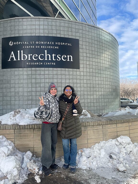

Milena Yalyzhko from Sumy State University, Ukraine, will be hosted by the University of Manitoba, Canada, for her secondment from 4th February 2025 to 3rd April 2025. During this period, she will focus on in vitro assessments of PCL-MXene nanomembranes in cardiovascular applications, specifically evaluating their biocompatibility in cell cultures and investigating the effects of electrical stimulation on stem cell differentiation into cardiomyocytes. She will also assess the impact of nanofiber alignment on cell bundle formation and organization while collaborating with local teams to align her project objectives with the goals of the ESCULAPE project.
Her training activities will include hands-on experience in advanced biomaterials and cardiac tissue engineering, particularly the fabrication of electrospun nanomembranes with MXene coatings and the use of in vitro models for cardiac cell studies. Milena will also enhance her skills in microscopy, viability assays, and molecular techniques for assessing cardiomyocyte markers, while benefiting from exposure to cutting-edge lab techniques and project management in the Horizon Europe framework.
From 8 February to 7 March 2025 Miglė Stančiauskaitė visited Biofabics LDA.
The main goal of this secondment was to explore the incorporation of MXenes into microfluidic devices and investigate the formation and characterization of MXene layers on 3D printing resin using spectroscopic ellipsometry (SE). Given Biofabics' expertise in 3D-printed microfluidics, the focus was on identifying strategies to enhance surface conductivity, wettability, and biosensing capabilities through MXene coatings.
A significant part of the secondment involved gathering information on MXene applications in microfluidics, reviewing relevant literature, and discussing potential approaches with colleagues at Biofabics. I also participated in internal meetings, gaining insight into ongoing projects at Biofabics and their expertise in bioprinting, biofabrication, and microfluidic system development. Additionally, I attended seminars on electrospinning, broadening my understanding of fabrication techniques.
During the secondment, simple 3D-printed structures were chosen, with a goal of coating them with MXenes and using SE to assess thickness, uniformity, and optical properties of the deposited layers. The findings will contribute to the development of MXene-enhanced microfluidic devices for biosensing applications, optimizing their performance for point-of-care diagnostics and analytical systems
Mr. Veeramuthu, representing RESPILON MEMBRANES S.R.O. in the Czech Republic, conducted an academic and collaborative visit to the University of Latvia from 8 February to 7 April 2025. The primary objective of the visit was to strengthen cooperation between industry and academia, with a focus on nanomaterials and advanced membrane technologies for biomedical applications.
During his stay, Mr. Veeramuthu was actively involved in research activities at the Laboratory of Advanced Biomaterials and Biophysics. He participated in scientific discussions, laboratory meetings, and seminars that facilitated knowledge exchange and joint planning of experimental work. As part of the collaborative research, 3D electrospun membranes were fabricated using chitosan both as a standalone biopolymer and in combination with other synthetic polymers such as polycaprolactone (PCL) and poly(ethylene oxide) (PEO). These blends aimed to optimize the mechanical strength, fiber uniformity, and biological functionality of the membranes for potential biomedical applications, particularly in wound healing and tissue engineering.
The electrospun membranes were subjected to structural analysis using scanning electron microscopy (SEM), which revealed key insights into fiber morphology, alignment, and surface architecture. The incorporation of PCL and PEO into the chitosan matrix resulted in improved fiber uniformity and interconnectivity, which are essential features for promoting cell adhesion and nutrient diffusion in biomedical scaffolds.
This collaboration not only contributed to ongoing experimental developments but also laid the groundwork for future joint research proposals, particularly in the areas of functional nanofiber-based membranes and advanced wound dressings. The visit concluded successfully with the intention to maintain long-term collaboration between RESPILON MEMBRANES S.R.O. and the University of Latvia, supporting innovation at the interface of material science and biomedicine.
During his secondment at Respilon from 16th February 2025 to 15th April 2025, Sergiy Kyrylenko focused on optimizing conditions for the industrial-scale synthesis of conductive nanofibrous polymeric membranes using MXenes with various properties and compositions. His work included the initial phase of coating electrospun polymeric membranes with surface layers of MXenes to impart electroconductivity. Sergiy also explored the dissolution of MXenes in polymer solutions, enabling their direct use in the electrospinning process.
As part of his project, he investigated the inclusion of other functional components, such as antibiotics, into the electrospinning solution to create nanofibrous membranes with specific biomedical properties.
Throughout his visit, Sergiy collaborated with Dr. Baturalp Yalçınkaya and Dr. Matej Buzgo at Respilon, engaging in detailed discussions on the technical aspects of electrochemical processes during electrospinning with conductive polymer solutions. The team also compared electrospinning protocols from various labs, including the University of Latvia, and discussed optimizing conditions for large-scale production of nanofibrous membranes using the multi-jet electrospinning strategy. These discussions were crucial for advancing the feasibility of producing industrial quantities of nanofibrous membranes with tailored properties.
Additionally, extensive brainstorming sessions were held to explore innovative methods for producing polymer nanomembranes with selective permeability, advancing the potential applications of the materials.
Sergiy was provided with specialized training in electrospinning techniques, particularly using the multi-jet strategy. Under the guidance of Respilon’s team, he learned how to assess electrospun nanomembranes using scanning electron microscopy (SEM) and light microscopy, which greatly enhanced his technical expertise. Sergiy expressed his appreciation for the warm hospitality and knowledge-sharing environment provided by Respilon’s R&D facility.
Matej Buzgo and Baturalp Yalcinkaya undertook a secondment from Respilon Membranes s.r.o. to Biofabics Lda, from 25 February 2025 to 6 March 2025 (Matej Buzgo) and 25 February 2025 to 1 April 2025 (Baturalp Yalcinkaya). During this period, they received practical training in laboratory safety and the operation of 3D bioprinting systems, including demonstrations of bio-ink handling and fundamental printing procedures. Initial experiments were conducted with simple geometries to evaluate parameters such as extrusion speed, temperature, and print resolution. Subsequently, more complex 3D structures were printed to investigate system performance in greater detail, considering the effects of layer height, nozzle movement, and bio-ink viscosity on structural stability. The outcomes were documented in a draft working protocol outlining key findings on bio-ink behaviour and 3D bioprinter efficiency, along with a step-by-step guide for future bioprinting activities
Two researchers from the Adam Mickiewicz University, Poznań, Poland (AMU), Andriy Lys and Irfan Hanif, spent a month at INSERM (Institut National de la Santé et de la Recherche Médicale) from 1st March to 30th March 2025. The visit was part of a collaborative effort aimed at deepening their knowledge of stem cell work and neurocardiac research and strengthening ties between the two research institutions.
As their secondment progressed, they were introduced to the intricate process of working with induced pluripotent stem cells (iPSCs). Andriy and Irfan learned how iPSCs are cultured and maintained, and observed the steps involved in directing these cells towards differentiation into both cardiomyocytes and neurons. Andriy and Irfan had the chance to observe calcium imaging experiments, where changes in intracellular calcium were visualized using fluorescent dyes. This experience gave them a deeper understanding of how researchers study cell function in real time, a concept they had previously encountered only in theoretical studies. Additionally, they observed multi-electrode array (MEA) recordings, which are used to measure the electrical activity of cells within a network.
The visit also provided ample opportunities for discussion with INSERM researchers, fostering a valuable exchange of knowledge and ideas. Andriy and Irfan are now better equipped with new skills in stem cell culture, live-cell imaging, and electrophysiological measurements, and are eager to apply this knowledge to their ongoing research at AMU.
During the period 03.03.2025 – 02.05.2025, Alexander Tamashevski from Adam Mickiewicz University (Poland) carried out a secondment at RESPILON MEMBRANES S.R.O. (Czech Republic).
The main objective of the secondment was to develop and characterize 3D porous membranes based on biocompatible and biodegradable synthetic polymer poly(ε-caprolactone) (PCL) using needle and needless electrospinning techniques. The obtained fibers were analysed using scanning electron microscopy (SEM) to evaluate morphology and structural uniformity. Needle electrospinning provided 3D scaffolds with more homogeneous fiber distribution. However, the pore size of these scaffolds was found to be too small for efficient cell penetration, thus limiting the diffusion of cells through the material. To overcome this limitation and enhance porosity, a modified electrospinning process with a chemical foaming agent was explored. This approach demonstrated potential for improving scaffold architecture and increasing the material’s suitability for tissue engineering applications.
The secondment allowed the participant to gain practical experience in electrospinning methods, SEM imaging, and structural analysis, as well as to collaborate with industrial researchers on optimizing polymer scaffold fabrication.
During the visit from March 14 to April 13, Igor Iatsunskyi was actively involved in research and development activities at Respilon, focusing on electrospinning techniques and nanofiber membrane fabrication. He gained practical experience operating both needle-based and needleless electrospinning equipment, with particular attention to adjusting critical process parameters such as solution feed rate, voltage, and needle-to-collector distance to control fiber morphology. Igor completed safety training relevant to high-voltage equipment and solvent handling, and worked closely with Respilon staff to troubleshoot typical challenges such as needle clogging, solution instability, and brittle fiber formation.
He contributed to the preparation of various polymer and composite solutions, especially those based on chitosan and PLA, and tested different solvent-to-polymer ratios to ensure electrospinning stability. His work included evaluating solutions' spinning behavior and analyzing the produced fibers' morphology and uniformity. Igor participated in quality control procedures, including SEM imaging and fiber diameter measurements, and assisted in characterizing membranes produced under both laboratory and industrial conditions. The visit also provided him with valuable insights into industrial-scale workflows, quality assurance protocols, and the practical application of electrospun membranes for biomedical use.
Matej Buzgo took part in secondments from Respilon Membranes s.r.o. to Biofabics Lda from 17 March to 1 April 2025 and 25 April to 31 May 2025. The main focus of these visits was the development of sophisticated 3D-printed organ-on-chip devices and bioreactor chambers with optimized geometries and functional properties for tissue engineering and 3D cell culture applications. The constructs were designed to include nanofibrous scaffolds reinforced with MXenes, aiming to enhance both mechanical strength and biocompatibility.
During the secondments, Matej received extensive training on the operation of 3D bioprinting systems and the fabrication of complex structures. The printed devices were assessed for mechanical stability and biological performance, emphasizing their potential integration with electrospun nanofibrous scaffolds.

Between March 25 and April 5, 2025, three researchers from the University of Latvia – Volodymyr Deineka, Martins Boroduskis, and Karīna Goluba – participated in a scientific secondment at BIOFABICS LDA, an innovative biotechnology company based in Porto, Portugal. This exchange was carried out within the framework of the Horizon Europe project “ESCULAPE,” which focuses on the development of advanced biomaterials and biofabrication technologies for regenerative medicine.
Volodymyr Deineka focused on mastering 3D bioprinting techniques using MXene-filled bioinks. The training involved printing both simple and complex 3D structures and evaluating their mechanical and biological properties. This work contributes to the development of smart, bioactive scaffolds for tissue engineering.
Martins Boroduskis worked on bioreactor systems, learning to set up and operate these platforms for the cultivation of cells on bioprinted scaffolds. During the first week, Martins configured bioreactor parameters and optimized environmental conditions. In the second week, he conducted experiments to assess cell proliferation and viability using biochemical and microscopy techniques.
Karīna Goluba concentrated on the design and optimization of microfluidic systems for tissue engineering applications. She began by designing and adapting microfluidic devices to support efficient nutrient and oxygen delivery for cell culture. In the second week, she tested and optimized these systems to improve performance in dynamic in vitro environments.
This secondment strengthened the collaboration between the University of Latvia and BIOFABICS, enabling hands-on knowledge transfer and interdisciplinary teamwork in advanced biofabrication. The newly gained expertise in 3D bioprinting, bioreactors, and microfluidics will be integrated into ongoing research activities and educational initiatives at the University of Latvia.
The team is planning to present the results of their work at international conferences and contribute to scientific publications in the fields of biomaterials and tissue engineering. The secondment has also laid the foundation for new collaborative research projects within the ESCULAPE consortium and beyond.
Matej Buzgo and Baturalp Yalcinkaya undertook secondments from Respilon Membranes s.r.o. to Biofabics Lda, spanning 4 April to 22 April 2025 for Matej Buzgo and 4 April to 31 May 2025 for Baturalp Yalcinkaya. These secondments focused on gaining in-depth understanding and practical experience in electrospinning techniques for the fabrication of polymeric fiber scaffolds engineered to support the adhesion, growth, and differentiation of human cells, with particular emphasis on neurons and cardiomyocytes.
During their stay, they collaborated closely with Biofabics researchers to explore the key parameters influencing fiber morphology, alignment, and density. They observed and participated in the setup and operation of electrospinning equipment, including preparation of polymer solutions, environmental control, and optimization of spinning conditions. Discussions were held on the selection of conductive and biocompatible polymers suitable for neural and cardiac tissue engineering applications.
During their visit to Adam Mickiewicz University (AMU) in Poznań, Poland, from April 13 to May 12, 2025, Ilia Yanko and Anton Roshchupkin from Sumy State University (SSU), Ukraine, made significant progress in applying Raman spectroscopy and atomic force microscopy (AFM) to the study of polycaprolactone (PCL) and chitosan electrospun membranes, both in their pristine form and modified with MXenes. The purpose of the visit was twofold: to undertake practical training and to advance the biological evaluation of MXene-integrated electrospun membranes.
By utilizing these techniques, they successfully explored the correlation between the membranes’ physical characteristics and chemical composition. Notably, Raman spectroscopy enabled the detection of changes in the membranes, MXene flakes, and cell responses during in vitro experiments. These results not only support previous findings but also lay the foundation for upcoming in vivo studies. Ultimately, the data may offer critical insights into the therapeutic efficacy of these electrospun membranes, promoting their future application in biomedical fields.

During the secondment period from April 14 to June 13, Katya Klimovich from Adam Mickiewicz University (AMU) completed training at the University of Latvia (LU). She gained practical experience in essential mammalian cell culture techniques, including cell passaging, seeding, counting, cryopreservation, and recovery. Katya also took part in several experiments aimed at evaluating the cytotoxicity of various MXene nanomaterials.
Through this work, she developed a deeper understanding of how advanced nanomaterials interact with biological systems and how their potential toxicity can be systematically assessed. This hands-on experience significantly enhanced her interdisciplinary research skills. The secondment notably broadened her technical expertise and deepened her understanding of MXene-based materials for biomedical applications.
During his secondment at INSERM, Montpellier, from 26 April 2025 to 25 May 2025, Sergiy Kyrylenko focused on investigating the biomedical properties of MXene-based electroconductive electrospun polymer membranes. His research explored how MXene-coated PCL membranes support the differentiation of induced pluripotent stem cells (iPS) into cardiomyocytes, fibroblasts, and neurons, and the formation of functional junctions between these cell types in biochip environments.
At INSERM, Sergiy was trained in the protocols for iPS differentiation, particularly into functional cardiomyocytes and fibroblasts. He also learned protocols for forming cardiomyocyte-containing beating embryoid bodies (EBs) and participated in the evaluation of their electrical and mechanical properties using microdevices developed in the host lab. In addition, Sergiy engaged in advanced electrophysiological studies on ryanodine receptors from a rat cardiac disease model. These studies led to the conclusion that optimal performance of cardiomyocyte bundles requires co-culture with fibroblasts derived from the same iPS line.
He also provided reciprocal training to INSERM staff and students on the fundamentals of MXenes and their biomedical potential. Knowledge exchange with Dr. Albano Meli and others has laid the foundation for future collaboration, including the planned provision of aligned MXene-coated nanofiber membranes from the University of Latvia and the Materials Research Center in Kyiv. The results of this secondment are expected to contribute to a broader project investigating the maturation and performance of iPS-derived cardiomyocytes and fibroblasts under various culture conditions.

Viktoriia Korniienko from the University of Latvia participated in an exchange visit to Vilnius University, Lithuania, from May 2nd to May 16th, 2025. The visit focused on surface modification of materials using MXene integration and structural and chemical characterization.
During the visit, samples were modified by incorporating MXene materials using a pressure-assisted infiltration method, ensuring uniform distribution. The samples were analyzed for changes in crystalline structure, surface functional groups, and chemical bonds. The results showed significant changes compared to unmodified samples, confirming successful MXene incorporation.
The findings lay the groundwork for future collaboration and applications of MXene-based surfaces in sensing materials. Future work will focus on evaluating the electrical conductivity of the MXene-modified samples for potential use in electroconductive and sensing materials.
Viktoriia Holobnycha from Sumy State University, Ukraine (May 16 – May 30) — conducted a research visit to the University of Eastern Finland (UEF).
The visit aimed to develop and validate an Electrical Impedance Tomography (EIT) protocol for assessing conductivity in MXene-coated scaffolds. This protocol was used on MXene-coated scaffolds, revealing significantly improved and more uniform conductivity. Results were discussed with UEF researchers, fostering future collaboration in advanced biomaterials.In addition to their experimental work, the visiting researchers delivered presentations at the UEF Institute of Public Health and Clinical Nutrition, introducing their ongoing scientific projects. She also participated in the MicroAmes test and other experimental activities in the Food Research Laboratory. The visit provided opportunities for hands-on laboratory engagement and constructive scientific dialogue, leading to the joint planning of future research and development initiatives in collaboration with Professor Marjukka Kolehmainen, Dr. Jenni Korhonen, and Professor Reijo Lappalainen, along with the Biomaterials Technology group at the Department of Technical Physics, University of Eastern Finland.
In May 2025, Viktoriia Korniienko from the University of Latvia (May 18 – June 1) conducted a research visit to the University of Eastern Finland (UEF) as part of a collaborative scientific exchange.
The primary objective of the visit was to develop and validate an Electrical Impedance Tomography (EIT) measurement protocol to assess conductivity distribution in MXene-coated and uncoated scaffolds. In the first phase, the EIT system was calibrated and tested using uncoated scaffolds to establish baseline conductivity values. In the second phase, the same protocol was applied to MXene-coated scaffolds to evaluate the impact of the coating on both local and overall conductivity. The results confirmed that MXene coating significantly enhanced scaffold conductivity and promoted a more uniform distribution of conductive regions. These findings were discussed with researchers at UEF, laying a foundation for future collaboration in the field of advanced biomaterials.
From June 2 to June 16, 2025, Senior Researcher Dr. Maksym Pogorielow, Head of the Advanced Biomaterials and Biophysics Laboratory Institute of Atomic Physics and Spectroscopy, Faculty of Science and Technology, together with Dr. Volodymyr Deineka, completed a successful scientific secondment at Vilnius University (Lithuania) within the framework of the Horizon Europe project ESCULAPE (“Electro-conductive polymeric 3D scaffolds as novel strategies for biomedical applications”).
The secondment focused on advanced materials characterization and aimed to evaluate the effect of MXene nanoparticles on the crystallinity and structural organization of electrospun polymer fibers. During their stay, the researchers carried out detailed X-ray diffraction (XRD) analyses of fiber samples prepared from polymer solutions with and without MXene additives. The main objective was to understand how the incorporation of MXenes influences crystalline phases, interlayer distances, and the structural stability of the polymer matrix.
The secondment also laid the groundwork for long-term collaboration between the participating institutions. The collected data will contribute to future joint scientific publications and conference presentations. This visit further supports the development of new project ideas focused on advanced nanomaterials for regenerative medicine and drug delivery. The research team expresses gratitude to their colleagues at Vilnius University for their warm welcome and valuable scientific exchange.

From July 7 to 21, Dr. Viktoriia Korniienko and Dr. Kateryna Diedkova from the University of Latvia conducted a research visit to Biofabric, Porto. The visit focused on the development of advanced 3D-printed organ-on-chip and bioreactor chambers with optimized shapes and functional properties suitable for tissue engineering and 3D cell culture. These systems were designed to incorporate nanofibrous scaffolds reinforced with MXenes, aiming to improve mechanical stability and biological compatibility. During the visit, the researchers received training in operating 3D bioprinting systems. These constructs were evaluated in terms of their mechanical integrity and biological performance, particularly focusing on their potential integration with electrospun nanofibrous scaffolds for tissue engineering applications. In parallel, the team worked on the design and optimization of microfluidic systems tailored for tissue engineering.
From 23 June 2025 to 3 July 2025, Albano Meli undertook a secondment from Inserm to Biofabics, focusing on observing and understanding the application of electrospinning techniques to fabricate engineered polymeric fiber surfaces designed to support the adhesion and growth of human cells, particularly neurons and cardiomyocytes. During his stay, Dr. Meli engaged with Biofabics researchers to explore the parameters influencing fiber morphology, alignment, and density. He observed the setup and operation of electrospinning equipment, including polymer solution preparation and environmental control, and took part in discussions on the selection of conductive and biocompatible polymers suitable for cardiac and neural applications. He also assessed the potential integration of fiber-based scaffolds into 3D bioprinting workflows for cardiac tissue engineering. The visit involved technical exchanges on scaffold characterization methods, such as mechanical testing, and knowledge-sharing sessions comparing approaches used at Inserm for 2D cardiac sheets and 3D cardiac organoid modeling with Biofabics’ electrospun constructs.
From June 25 to July 24, Arvīds Borovskis from the University of Latvia completed a secondment at BIOFABICS (Portugal), focusing on technical training in the use of 3D bioprinted organ-on-chip systems.
During the secondment, the participant received hands-on training in laboratory safety and the operation of 3D bioprinting systems. The training included practical demonstrations of bio-ink handling and basic printing protocols. Initial testing was performed using simple geometries to assess parameters such as extrusion speed, temperature, and print resolution. This was followed by the printing of more complex 3D structures to analyze system performance in greater depth, including the influence of layer height, nozzle movement, and bio-ink viscosity on structural stability.
The results were compiled into a draft working protocol documenting key findings on the behavior of bio-inks and the effectiveness of the 3D bioprinting system. A step-by-step guide was developed for future bioprinting procedures.
Dace Gulbe undertook a secondment within the framework of the ESCULAPE project, from Latvijas Universitāte (LU), Latvia to BIOFABICS LDA, Portugal, from 09.07.2025 to 23.07.2025. The purpose of this secondment was to enhance skills in the commercialization of research results, including intellectual property protection, patent application preparation, and research commercialization. The knowledge gained will be applied to the effective management of the ESCULAPE project, improving result dissemination strategies, engaging stakeholders, and communicating scientific achievements.


Klara Kuzņecova, a researcher from the Institute of Electronics and Computer Science (EDI), Latvia, carried out a two-month secondment at the Adam Mickiewicz University in Poznań (AMU), Poland, from 14 July to 13 September 2025, within the framework of the ESCULAPE project.
The purpose of the visit was to conduct experimental research on the evaluation and optimization of MXene (Ti₃C₂Tₓ) dispersions and coatings for flexible bioelectrodes aimed at reducing skin-electrode contact impedance and minimizing motion artefacts. During the secondment, Ms Kuzņecova prepared and characterised MXene dispersions, deposited them on flexible substrates, and developed a coating series with controlled thickness and binder composition. In addition, she performed interface engineering using hydrogel and electrospun PLA fiber interlayers to improve coating adhesion and stability. A comprehensive set of electrical and microstructural characterizations was completed, including four-point probe surface resistance measurements, bioimpedance spectroscopy across a wide frequency range, and electrochemical stability evaluation.
The results of the secondment will be applied at EDI for further prototype integration, impedance and stability testing on wearable systems, and follow-up publications. The collaboration strengthened methodological and technical capacity in both institutions and established a solid basis for continued cooperation, data exchange, and future student mobility under the ESCULAPE project.
From July 20 to August 19, 2025, Senior Researcher Dr. Maksym Pogorielow, Head of the Advanced Biomaterials and Biophysics Laboratory, Institute of Atomic Physics and Spectroscopy, Faculty of Science and Technology, together with Dr. Volodymyr Deineka, completed a successful one-month scientific secondment at RESPILON MEMBRANES S.R.O. (Czech Republic) within the framework of the Horizon Europe project ESCULAPE (Electro-conductive polymeric 3D scaffolds as novel strategies for biomedical applications).
The secondment was dedicated to advancing electrospinning technologies and optimizing MXene nanomaterial integration into electrospun fibers. During the stay, the researchers underwent specialized training in laboratory safety and electrospinning equipment operation. They then worked on the development of MXene–polymer solutions, tested electrospinning parameters, and evaluated the influence of MXene incorporation on fiber morphology and conductivity. In the final stage, a reproducible MXene integration protocol was established, including quality control steps and reproducibility assessments of the obtained nanofibers.
This scientific visit provided valuable expertise and new methodological approaches for the development of MXene-based nanocomposites in biomedical applications, particularly in tissue engineering and drug delivery systems. It also reinforced collaboration between the University of Latvia and RESPILON MEMBRANES S.R.O., laying the foundation for future joint projects, conference presentations, and scientific publications. The research team expresses its sincere gratitude to the colleagues at RESPILON MEMBRANES S.R.O. for their professional support and constructive scientific exchange.
In July–August 2025 three researchers from Adam Mickiewicz University (Poland), Andrii Lys, Dr. Maksim Makhavikou, and Dr. Alexander Tamashevski, took one-month research secondments at BIOFABICS, a leading company in 3D bioprinting and organ-on-chip technologies based in Porto, Portugal. The visits aimed to provide hands-on experience in the design and fabrication of bioreactor chambers and microfluidic organ-on-chip systems using high-resolution SLA 3D printing. The researchers focused on developing customized microfluidic platforms for in vitro testing of nanofiber and 3D porous scaffolds in cardiac and neural tissue models.
These activities will support the optimization of scaffold–cell interactions, enhance the integration of 3D-printed systems into tissue engineering workflows, and strengthen collaboration between AMU and BIOFABICS. The secondments will also promote knowledge exchange and joint development of advanced biofabrication and testing platforms for future regenerative medicine applications
From July 25 to August 24, Arvids Borovskis from the University of Latvia completed a secondment at Respilon (Czech Republic), focusing on advanced training in electrospinning technologies.
During the visit, he received practical training on the safe use of electrospinning equipment and explored a range of fiber production techniques, including both horizontal and vertical configurations. He conducted experimental work using MXene-enriched polymer solutions to produce nanostructured, electrically conductive fibers for potential biomedical applications. The work included optimizing electrospinning parameters and analyzing the impact of viscosity and conductivity on fiber morphology.
By the end of the secondment, detailed experimental protocols were developed, along with recommendations for future research. The experience contributed significantly to strengthening cooperation between the University of Latvia and industry partners in the field of advanced nanomaterials and biomaterials.
From July 29 to August 28, Dr. Kateryna Diedkova from the University of Latvia carried out a research visit to RESPILON MEMBRANES S.R.O. in Prague, Czech Republic.
The visit was dedicated to the development of advanced 3D electrospun porous scaffolds with conductive properties for biomedical applications in regenerative medicine. These tissue-engineering platforms were fabricated using conventional electrospinning, combined with the incorporation of MXenes, with the aim of enhancing their electroconductive performance.
During the visit, Dr. Diedkova also received training in innovative fiber-spinning technologies, including coaxial and multijet electrospinning. In parallel, collaborative work was carried out on the design and optimization of electrically conductive 3D porous scaffolds with predictable properties, integrating 2D nanomaterials for tissue-engineering applications
From August 4 to August 18, 2025, Viktoriia Korniienko from the University of Latvia (LU) participated in a research exchange at Vilnius University, Lithuania.
The aim of the visit was to carry out surface modification using MXene materials and to analyze the resulting structural and chemical changes in comparison with unmodified samples. Advanced analytical techniques were employed, including X-ray diffraction (XRD), Fourier-transform infrared spectroscopy (FTIR), and Raman spectroscopy.
During the exchange, sample preparation and MXene surface modification were performed. Initial structural and chemical analyses were conducted using XRD and FTIR. In the second week, detailed Raman spectroscopy was carried out, along with additional XRD and FTIR measurements to obtain in-depth information on chemical bonding and functional group transformations. The results confirmed changes in chemical structures compared to unmodified samples and helped define directions for future research.
Anton Roshchupkin (from September 5 to September 20, 2025) and Oleksandr Solodovnyk (from September 6 to September 20, 2025) from Sumy State University (SSU) completed a secondment at RESPILON in the Czech Republic. The main objective of their research activities was to develop electrospun membranes with electroconductive properties and to analyse their physical and mechanical characteristics.
During the secondment, they participated in the fabrication of electroconductive electrospun membranes and used specialized equipment to evaluate their mechanical strength and physical performance. Scanning electron microscopy (SEM) was employed to assess the surface topography of the produced membranes. They also engaged in discussions regarding the potential applications of these materials as 3D scaffolds for biomedical purposes.
Both researchers received practical training in essential aspects of the electrospinning process, including the preparation of polymer solutions, as well as the calibration and maintenance of the electrospinning equipment. This experience enhanced their expertise in electrospinning techniques and material characterization methods.
The collaboration facilitated the establishment of new professional connections, opening opportunities for future joint research. The knowledge and skills gained during this secondment will be shared at SSU through internal workshops, training sessions, and collaborative seminars aimed at strengthening the institutional expertise in electrospinning and advancing interdisciplinary research.


From 6 September to 5 October 2025, Andrii Liutyi visited Respilon Membranes s.r.o. in Prague as part of the ESCULAPE project. During the secondment, he participated as a visiting researcher, focusing on the development and optimization of nanofibrous membranes for biomedical applications. The research activities included electrospinning of polymeric fibers, investigating how process parameters affect fiber morphology, mechanical properties, and overall scaffold quality.
Special attention was given to the integration of conductive nanomaterials, such as MXenes, into the membranes to enhance electrical conductivity and biocompatibility. Andrii also explored surface functionalization techniques to improve cell adhesion on scaffold surfaces. Hands-on training included material characterization methods, such as microscopy-based morphological analysis, mechanical testing, and porosity assessment.
Collaboration with Respilon’s R&D team provided insights into industrial workflows, process standardization, and quality control procedures. Discussions and technical sessions helped align laboratory research with industrial production standards. The secondment offered practical experience in scaling up nanofiber fabrication and optimizing scaffold composition for biomedical applications.
Overall, the visit strengthened expertise in electro-conductive scaffold development, provided valuable industrial perspectives, and contributed to the ESCULAPE project’s goal of creating advanced biomaterials for tissue engineering


During a one-month research secondment (16.09.2025-15.10.2025), Aleksandrs Dutovs from the University of Latvia explored innovative materials and advanced analytical techniques in Adam Mickiewicz University (AMU) in Poznań, Poland.
The researcher received hands-on training in state-of-the-art Fourier Transform Infrared (FTIR) and Raman spectroscopy - powerful tools for studying the structure of modern materials. Polymer and MXene-based composite samples were prepared and coated, followed by visual inspection and evaluation to assess coating uniformity and surface features. Detailed morphology analysis was performed using a Scanning Electron Microscope (SEM), providing high-resolution insights into the structure and distribution of MXene layers within the polymer matrix. The work resulted in valuable insights into the chemical composition and structural behaviour of these next-generation materials.
Through close collaboration and knowledge exchange, the secondment fostered innovation, strengthened international research links, and contributed to the advancement of sustainable material technologies.
Serhii Dmytruk from Sumy State University (SSU), Ukraine, completed a secondment at RESPILON s.r.o. in the Czech Republic from September 20 to October 19, 2025, as part of the Horizon Europe MSCA-SE project ‘Electrically Conducting Polymer 3D Scaffolds as New Strategies for Biomedical Applications’.
The main goal of this secondment was to participate in research activities, focusing on the design and synthesis of electrically conductive electrospun membranes using both traditional and multi-needle electrospinning techniques. The experimental work aimed to develop 3D mats based on PLA and PCL polymers with controlled structural parameters such as fibre diameter, length, thickness, and porosity. Special attention was given to optimizing electrospinning conditions to achieve uniform fibre morphology and desired electrical conductivity.
During the secondment, Serhii gained practical experience in preparing and optimizing polymer solutions and mixtures (PLA, PCL, and their composites) suitable for electrospinning, including the adjustment of viscosity, conductivity, and solvent ratios. He studied key process parameters for obtaining uniform nanofibers and 3D scaffolds with predefined morphology and mechanical properties
Baiba Zandersone (University of Latvia) conducted a research visit to the University of Eastern Finland (UEF) from 28 September to 12 October as part of a collaborative scientific exchange. The main objective of the visit was to develop and validate an Electrical Impedance Tomography (EIT) measurement protocol for assessing the conductivity distribution in MXene-coated scaffolds.
During the visit, the EIT system was calibrated, and measurements were performed to establish reference conductivity maps. The validated protocol was subsequently applied to MXene-coated scaffolds prepared using the membrane-deposition method.
The results revealed that the MXene coating had a pronounced effect on scaffold conductivity. The extent of this influence depended on the deposition method, coating thickness, and uniformity, resulting in a more homogeneous distribution of conductive regions across the scaffold.
The obtained data were analysed and discussed with researchers from the host institution, fostering knowledge exchange on the electrical properties of MXene-based biomaterials and strengthening the basis for future collaborative research in this field.
Miglė Stančiauskaitė (Vilnius University, VU) completed a secondment to Biofabics from 30 September to 29 October 2025 as part of the ESCULAPE project.
The main goal of this secondment was to explore how MXenes can be incorporated into microfluidic devices, with emphasis on forming and characterizing MXene layers on 3D-printed resin and glass substrates using spectroscopic ellipsometry (SE). BioFabics’ expertise in 3D-printed microfluidics allowed to focus on strategies to improve surface conductivity, wettability, and readiness for biosensing through MXene coatings.
Literature review of MXene coatings for polymer and glass used in microfluidic systems was done, alongside processing and interpreting experimental data brought from my home institution. I also participated in internal meetings to understand BioFabics’ ongoing fabrication work and constraints. During an internal presentation, I shared my experience with QCM-D and SE, highlighting how these techniques assess surfaces and surface modifications and can support quality control of conductive coatings. Initial, small-scale trials with drop-casting MXene dispersions onto flat resin surfaces were conducted to probe interactions and compatibility of untreated resin with aqueous MXene solutions across concentrations.
The outcomes include a preliminary resin and glass-compatible MXene deposition strategy, along with identified risks such as oxidation and delamination under flow. These findings will guide next steps at my home institution, validating adhesion and continuity via QCM-D and SE and assessing stability in flow conditions.
From 30th September to 29th October 2025, the secondee, Marina Sapauskiene, visited Biofabics for a one-month secondment. The main goal of the secondment was the fabrication of polymeric nanofibers using the electrospinning technique, followed by their coating with MXene solutions.
During the secondment, polymeric nanofibers were produced under various electrospinning conditions to optimize fiber morphology and uniformity. The resulting nanofibers were subsequently coated with MXene solutions prepared in different concentrations and solvent systems to enhance their electrical and surface properties. Additional electrical and mechanical tests were carried out to assess the effects of MXene deposition and the applied optimization parameters on nanofiber performance. These activities provided valuable hands-on experience in nanofiber fabrication, surface modification, and advanced material characterization, contributing to the development of high-performance MXene-coated polymeric nanofibers.
Future research will focus on refining electrospinning and coating techniques for MXene-modified nanofibers. Planned work includes optimizing material properties, improving coating adhesion, and exploring new polymer–MXene combinations for biomedical and functional applications. The knowledge and experimental skills gained during the Biofabics secondment will serve as a foundation for continued development of MXene-coated polymeric nanofibers with enhanced performance for potential advanced applications

From October 1 to October 31, 2025, a month-long secondment to RESPILON Membranes s.r.o. in Prague, Czech Republic, was carried out by Viktor Kulibaba and Viktoriia Kazban, researchers at the Center for Collective Use of Scientific Equipment “Biomedical Research Center” of Sumy State University. The purpose of the visit was to participate in the design and synthesis of electrically conductive electrospun membranes using MXenes of various compositions within the framework of the project “Electro-conductive polymeric 3D scaffolds as novel strategies for biomedical applications.”
During the secondment, the researchers worked on developing 3D mats with controlled structural parameters, such as fiber diameter, length, orientation, thickness, and overall dimensions, based on PLA and PCL polymers. The work involved the use of traditional, needle-free, and multi-needle electrospinning techniques to produce electroconductive scaffolds with tunable properties.
The skills and expertise gained during this visit will be applied in future research activities focused on developing advanced 3D mat-based components incorporating MXenes for biomedical and tissue engineering applications
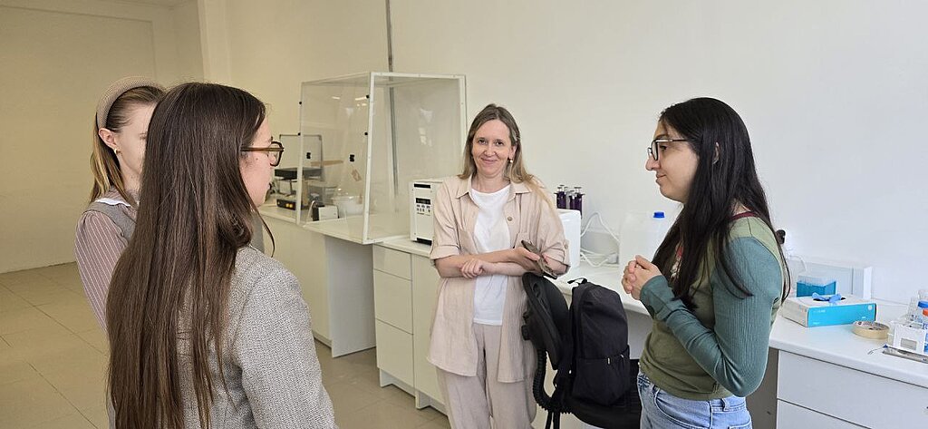
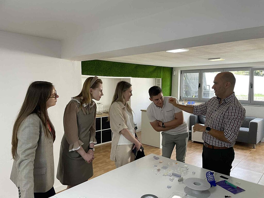

Two representatives of Sumy State University (Inna Chorna and Ilia Yanko) have completed a month-long secondment (01.10.2025-31.10.2025) at Biofabics Lda (Porto, Portugal) as part of the Horizon Europe MSCASE project “Electro-conductive polymeric 3D scaffolds as novel strategies for biomedical applications” (ESCULAPE). During their stay, they had the opportunity to delve into the exciting world of organ-on-chip technologies and 3D biotissue analogues working alongside the amazing Biofabics team. Their work focused on the development of 3D-printed organ-on-chip systems and bioreactors, exploring how advanced materials such as MXenes can improve tissue engineering and disease treatment. Beyond the lab, it was an incredible opportunity to exchange knowledge, share ideas, and see how engineering and biology come together to solve real biomedical issues
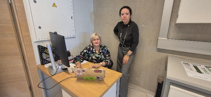

Almira Ramanaviciene (Vilnius University, VU) successfully completed a secondment to Latvia University (LU) from 5 to 19 October 2025 as part of the ESCULAPE project.
During the secondment, Prof. A. Ramanaviciene was focused on the deposition of MXene on the carbon substrate by two methods in order to obtained stable layer with the highest surface density and conductivity. Much attention was paid to the deposition of Ti3C2Tx MXene using drop casting method. It was observed that layering of MXenes on a surface did not yield the expected results. After optimization of MXenes concentration in the drop casting solution and deposition protocol, the formed MXene layer was characterized by electrochemical methods. It was observed that at optimized concentrations of MXene the registered current significantly increased due to the superior MXenes electrical properties.
In addition to experimental work, the importance of the MXenes biocompatibility and possible biocompatibility tests were discussed. The secondee at the host research institution received a theoretical training on the nanomaterial’s biocompatibility tests. This training significantly enhanced knowledge’s and formed the basis for practical experiments with MXene


Viktoriia Korniienko (University of Latvia) successfully completed a research visit to Vilnius University (VU) from 13 to 27 October 2025 as part of the ESCULAPE project.
During the visit, Dr. Korniienko evaluated how MXene materials influence the chemical and structural properties of modified surfaces using XRD, Raman, and FTIR spectroscopy. She prepared MXene-coated samples, ensuring uniform coating and accurate labeling for subsequent analysis.
The obtained results revealed that MXene coatings alter the surface chemical composition, promote the formation of functional groups, and induce structural changes, enhancing the materials’ electrical and functional properties. These findings highlight the potential of MXene-modified materials for future biomedical applications.